en

What are Radiological Examinations?

Radiological examinations in dentistry are an essential diagnostic tool that allows dentists to visualize structures that are not visible to the naked eye. They help in detecting and accurately diagnosing various problems related to teeth, jaws, and surrounding tissues. These examinations are also crucial for planning complex treatment procedures, such as implantation, orthodontic treatment, endodontic therapy, or surgical interventions.

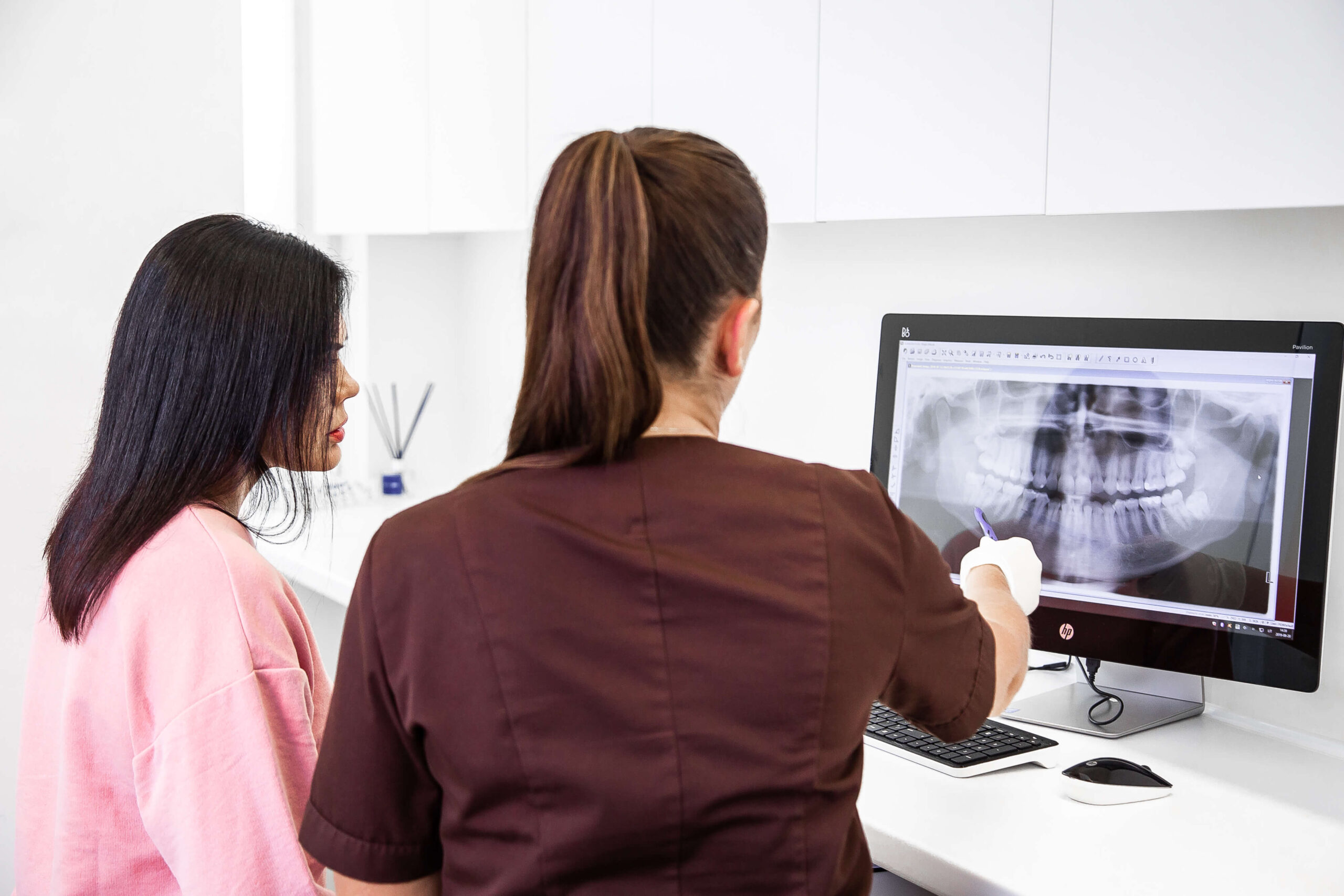
Radiological exams assess the patient’s oral and dental condition, enabling the dentist to create an accurate treatment plan. At CLINIC | DPC, only digital radiographs are used, which display the image on a computer screen and reduce radiation exposure by up to 90% compared to older X-ray machines.

Starting from

9€

Benefits of Radiological Examinations

Accurate Diagnosis

These examinations allow the dentist to accurately diagnose issues like cavities, periodontitis, cysts, tumors, root infections, and more.

Early Detection

X-rays help identify problems early, even before the patient experiences symptoms.

Treatment Planning

Essential for planning procedures like implantation, orthodontics, surgical procedures, endodontics, and other complex treatments.

Types of Radiological Examinations Performed at CLINIC | DPC

Panoramic X-Rays

Radiological exams capture the entire oral cavity in one image, including the upper and lower jaw, all teeth, sinuses, and the temporomandibular joint. They are used to assess tooth alignment, identify cysts, tumors, eruption issues, jaw fractures, and other conditions.

A panoramic dental X-ray is an essential diagnostic tool that allows dentists to thoroughly evaluate the patient’s oral health, diagnose various pathologies, and precisely plan treatment. It is widely used due to its convenience, speed, and ability to capture a comprehensive image of the entire oral area in one frame.

Dental X-Rays

A dental X-ray is a radiological test designed to give a detailed view of teeth, gums, surrounding tissues, and jaws. These images help dentists diagnose a wide range of oral health problems that might not be visible during a routine examination.

Dental X-rays show the entire tooth, from the crown to the root tip, and the surrounding bone. This is particularly useful for diagnosing root canal infections, cysts, and assessing bone structure.

Computed Tomography (3D CT)

Dental Computed Tomography (CT) is an advanced radiological test that provides a three-dimensional (3D) view of the teeth, jaws, and surrounding tissues. This examination allows dentists to accurately diagnose and plan complex treatment procedures.

Unlike traditional two-dimensional (2D) X-rays, CT scans create a detailed 3D model. These highly detailed images make it possible to precisely assess tooth root structure, bone density, nerve pathways, and other important anatomical features.

When 3D CT Scans are Typically Used

  • Before dental implant placement.
  • Before orthodontic treatment.
  • Before surgical procedures.
  • Before root canal treatment
  • After dental trauma.
  • Before orthognathic surgery.

Frequently Asked Questions

  • 01

    Are X-ray diagnostic tests safe?

    X-ray diagnostic tests do not cause any obvious negative effects on the body, as the exposure to radiation is very low. X-rays are performed only with a referral or during a consultation at the clinic.

Pay in installments 0%

We offer our patients the possibility of financing their treatment in a quick and simple way, in the form of convenient installments 0%.

Still have questions?

Whether you have questions about procedure, waiting time or compatibility with your insurance - call us and we will help you!